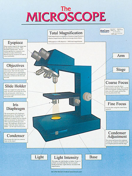
18 " by 24" (45.7cm by 61 cm)
Laminated for durability
State-of-the-art photography and illustration
wc01 - The Microscope
wc02 - Microscope Magnif.
wc03 - Microscope Focusing
wc04 - Bacteria
wc05 - Blood Cells
wc06 - Blood Typing
wc07 - Cells
wc08 - Mitosis
wc09 - Meiosis
wc10 - Epithelial Tissue
wc11 - Connective Tissue
wc12 - Bone Tissue
wc13 - Muscle Tissue
wc14 - Nervous Tissue
wc15 - Female Reproduction
wc16 - Male Reproduction
wc17 - Digestive System
wc18 - Urinary System
wc19 - Integumentary System
wc20 - Respiratory System
wc21 - Root Histology
wc22 - Stem Histology
wc23 - Leaf Histology
wc24 - Zygomycetes
wc25 - Ascomycetes
wc26 - Basidiomycetes
wc27 - Lily Life Cycle
wc28 - Fern Life Cycle
wc29 - Moss Life Cycle
wc30 - Pine Life Cycle
wc31 - Drosophila
wc32 - Pond I
wc33 - Pond II
wc34 - Pond III
wc35 - Porifera
wc36 - Cnidaria
wc37 - Mollusca
wc38 - Echinodermata
wc39 - Arthropoda
wc40 - Platyhelminthes
wc41 - Nematoda
wc42 - Annelida
wc43 - Chordata

wc44 - Floral Diversity
wc45 - Fleshy Fruits
wc46 - Dry Fruits
wc47 - HIV
wc48 - Animal Cell
wc49 - Plant Cell
wc50 - Bacterial Cell
wc51 - Cell Membrane
wc52 - Cell Wall
wc53 - Waterborne Pathogens
wc54 - Microbial Food Poisoning
wc55 - Virus I
wc56 - Virus II
wc57 - Virus III
wc58 - Amoeba
wc59 - Paramecium
wc60 - DNA
wc61 - Immune I
wc62 - Immune II
wc63 - Immune III
wc64 - Genetics 1
wc65 - Genetics 2
wc66 - Genetics 3
wc67 - Euglena
wc68 - The Flower
wc69 - Volvox
wc70 - Osmosis
wc71 - pH
wc72 - Photosynthesis